







Building upon the "Mechanics & Living Systems" operation (an initiative bringing together the researchers interested in biomechanics on the École Polytechnique campus)'s own seminar series, the Paris-Saclay University Biomechanics Seminar Series aims at structuring the biomechanics community of the Saclay plateau. It is organized jointly by Prof. Abdul Barakat, Dr. Dominique Chapelle, and Dr. Martin Genet. It is held tentatively on the last Thursday of each month at 14h, in the Sophie Germain amphitheater (map) of the Turing building (directions).
You can subscribe to the seminar series announcement mailing list here. Also, you can subscribe to the seminar series shared calendar using this link (do not download and import the calendar, simply create a new remote calendar with this address in order to always get the most up-to-date version).
For those especially interested in biomechanics/biophysics, please check out the Physics-Biology interface seminar organised at Université Paris Sud.
June 27, 2019: Prof. Giuseppe Rosi (Assistant Professor, Paris-Est Créteil Val-de Marne University and Multiscale Modeling and Simulation Laboratory (Paris-Est Marne-la-Vallée University/Paris-Est Créteil Val-de Marne University/CNRS), Créteil)
Waves and generalised continua in bone biomechanics and tissue engineering
Wave propagation in microstructured solids, such as living or artificial tissues, is a challenging topic in mechanics. In general, when the frequency of a dynamic sollicitation increases, the microstructure starts playing a role in the dynamic balance. In this case non-standard macroscopic effects emerge, the relationship between frequency and wavenumber becomes nonlinear and the medium is said to be dispersive. In this talk it will be shown that generalised continua (such as micromorphic or strain gradient) can be used to predict the accurate dynamic response in a frequency-wavenumber domain larger than that of the classic continuum. Examples will be given for microstructures based on honeycomb and gyroid unit cells. Finally, some perspectives on the design of artificial tissues with an enhanced ultrasound response will be discussed.
May 23, 2019: Prof. Christophe Clanet (CNRS Research Director, LadHyX (École Polytechnique/CNRS), Palaiseau)
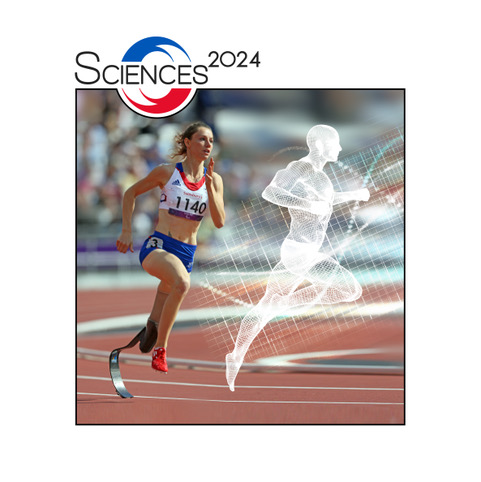 |
Physique du Sport et Sciences 2024 La physique consiste à observer le monde qui nous entoure et à chercher des lois permettant de rendre compte de nos observations. Dans cette conférence, le monde se limitera aux terrains de sports et l'on discutera de la physique associée à plusieurs sports olympiques et paralympiques dont le saut en longueur paralympique, l'aviron, la natation et le tir à l'arc. Dans un second temps, j'essayerai de convaincre quelques chercheurs de rejoindre Sciences 2024 pour épauler les équipes de FRANCE. Pour cela, je m'appuierai sur quelques exemples des besoins qu'elles expriment: optimisation des lames de course et de saut paralympique, oxymétrie embarquée, ... |
April 25, 2019: Dr. Anne-Virginie Salsac (CNRS Research Director, Biomechanics and Bioengineering Laboratory, UTC Compiègne)
Fluid structure interaction of a microcapsule in flow: application to the characterization, enrichment and sorting of capsule suspensions
Capsules consisting of a liquid droplet enclosed by a thin membrane are commonly encountered in nature in the form of cells, and in industrial processes (for instance to encapsulate active substances for targeted release). The wall mechanical properties are essential to guarantee the particle integrity and the release of the internal contents when and where necessary. However, when the capsules have a diameter of the order of a few tens of micrometers or less, it is very difficult to assess the mechanical properties of the membrane.
Modification of cell rigidity can, furthermore, be the consequence of diseases like cancers. Sorting cells based on their rigidity open interesting perspectives to differentiate between two cell populations, since conventional biological methods are known to be expensive. So far, most of the methods that have been designed to sort rare blood cells use the cell density or size as sorting parameters. We will see that microfluidic approaches can not only be used to characterize the mechanical properties of deformable capsules, but also sort them upon their rigidity or increase enrich suspensions.
I will briefly present how the three-dimensional fluid-structure interactions may be modeled in small to low inertial regimes and mostly detail the application of numerical simulations and microfluidic experimentations to characterize, enrich and sort suspensions.
February 21, 2019: Dr. Pascal Hersen (CNRS Research Director, Matter and Complex Systems Laboratory (Paris-Diderot University/CNRS), Paris)
Cybergenetics : control of gene expression and biological circuits
Gene expression plays a central role in the orchestration of cellular processes. We recently developed an experimental platform for real-time, closed-loop control of gene expression that integrates microscopy for monitoring gene expression in live cells, microfluidics to manipulate the cells environment, and dedicated software for automated imaging, quantification and model predictive control strategy. This method implements a dynamic interaction between cells and a computer, making it possible to control precisely the level of expression of a gene for both time-constant and time-varying target profiles, at the population level, and even at the single-cell level. I will discuss recent developments of this method and its relevance for systems and synthetic biology.
January 31, 2019: Prof. Rachele Allena (Assistant Professor, Georges Charpak Institute of Human Biomechanics, ENSAM-Paris/Paris XIII University)
From bone to cells and back: a journey through the mechanical environment
Most of the pathologies such as cancer or osteoporosis show their deleterious effects at the scale of tissues and organs. Similarly, when a prosthesis, a scaffold or a biomaterial is implanted in the body, one may not notice the bad functioning of the system until serious consequences appear at the macroscale. Nonetheless, the origin of such outcomes and behaviors must be looked for at the scale of the cell. It is in fact at the microscale, if not below, that cells interact with their environment and respond to different cues modifying the microstructure. Therefore, it is necessary to identify and quantify such processes in order to prevent specific pathologies and properly design biomechanical implants. In this talk I will focus on the bone remodeling process which takes place in both physiological and non- physiological conditions. I will present some interesting numerical models allowing to analyze fundamental cellular functions such as migration, which is critical during remodeling and may be highly influenced by the mechanical properties of the surroundings as well as by its morphology. These models, together with an accurate description of the bone microstructure, enable to consistently simulate the bone remodeling process and its initiation, which is assumed to be triggered by microcracks.
December 20, 2018: Dr. Olivier Theodoly (CNRS Research Director and Head, Adhesion & Inflammation Lab (INSERM/CNRS/AMU), Luminy Marseille)
Mechanics in immune cell recruitment
Immune cells, or leukocytes, are responsible for the main defense of our organism against infections. They continuously patrol the whole body via the blood system, until they eventually escape blood vessels and reach an inflamed region. This recruitment follows a coordinated sequence of events, including specific adhesion to inflamed vessels and directed migration within tissues. These sophisticated functions, which involve complex intracellular biochemical programs, rely also on passive mechanical properties. We will present here the role of cell rheology in a massive inflammatory disease (ARDS), and the functioning of cell guiding versus a hydrodynamic flow (mimicking blood flow) or versus gradients of substrate adhesion (mimicking blood vessels walls).
November 29, 2018: Dr. Irène Vignon-Clementel (Research Director, Inria & Sorbonne University, Paris)
Hemodynamics modelling of the local and global circulation: biomechanics and clinical application examples
Blood flow modelling builds on fluid mechanics principles in conduits, possibly in interaction with a solid wall. Depending on the biomechanics phenomena of interest to answer a medical question, the model represents local, 3D phenomena, or is reduced to 1D Euler equations or even 0D lumped model of the circulation to capture interactions between its different components. In any case, patient-specific or at least pathophysiologically relevant pressure and flow information needs to be integrated into such models. We will illustrate how such modelling can help to better understand congenital heart disease alleviation, the interaction of multiple components of such pathology and the mechanisms at play in partial ablation of liver. Vascular adaptation is an important component of the model to be predictive.
November 6, 2018: Prof. Arezki Boudaoud (Professor, Plant Reproduction and Development Institute, ENS-Lyon/CNRS/UCBL/INRA/INRIA)
Mechanical heterogeneities in plant morphogenesis
The two hands of most humans almost superimpose. Similarly, flowers of an individual plant have almost identical shapes and sizes. This robustness is in striking contrast with growth and deformation of cells during organ morphogenesis, which feature considerable variations in space and in time, raising the question of how organs and organisms reach well-defined size and shape. Because heterogeneous growth induces mechanical stress in tissues, we are exploring the biomechanical basis of morphogenesis. By combining approaches from developmental biology (molecular genetics, live imaging), and mechanics/physics (mechanical measurements, models), we are unravelling unexpected cellular patterns and behaviours. During this talk, I will discuss some of our recent results, aiming at a general audience.
October 25, 2018: Dr. Cécile Sykes (Team Leader, Institut Curie, Paris)
Tension build-up and membrane deformations by actin, studied with biomimetic systems
In order to unveil generic mechanisms of cell movements and shape changes, we design stripped-down experimental systems that reproduce cellular behaviours in simplified conditions, using liposome membranes on which cytoskeleton dynamics are reconstituted. Such stripped-down systems allow for a controlled study of the physical mechanisms that underlie cell movements and cell shape changes. Moreover, we are able to address biological issues within a controlled, simplified environment.
We reconstitute the actin cortex of cells at the membrane of liposomes, and characterize their mechanical properties. We will show how these cortices contract in the presence of myosin motors. A method using liposome doublets allows us to quantify cell tension change during contraction.
We reconstitute membrane tubules and spikes pulled or pushed by actin polymerization, which reproduce the formation of endocytic vesicles and dentritic filopodia. We will show how membrane tension and actin mechanical properties lead to the formation of inward or outward membrane deformations.
September 27, 2018: Prof. Bernard Sapoval (CNRS Research Director, Condensed Matter Physics Laboratory, École Polytechnique/CNRS, Palaiseau)
Gas exchanges in the human lungs
The understanding of gas exchanges and their pathological perturbations is a main object of pulmonary physiology. The gases of interest are the respiratory gases, oxygen O2 and carbon dioxide CO2 and gases used for diagnostic like carbon monoxide CO. And, because of their medical importance, the understanding of gas capture has been since the early days a major theme for experimental and conceptual modelisation efforts. Respiration being a complex phenomenon, there has been historically a continuous backwards and forwards motion between in vivo experimental data and modelisation.
We will discuss briefly three topics in which physics has brought new concepts in lung physiology.
1- Theory of oxygen capture. The role of heterogeneity.
2- Application to the prediction of the post-operative respiratory performances of patients that are candidates to pneumonectomy.
3- Theory of CO capture.
June 28, 2018: Dr. Cyprien Gay (CNRS Researcher, Laboratoire Matière et Systèmes Complexes, Université Paris Diderot)
Mechanical modeling of a developing biological tissue as both continuous and cellular: theory and (some) simulation tests
Deformability, growth, plasticity, relaxation: some of these ingredients span from the cytoskeleton to the tissue lengthscale. We will review arguments that can help us combine them in order to reproduce some features of a soft, active material, namely a developing animal tissue. We will thus progressively build a paradoxical formulation of the mechanical behaviour of the tissue as a continuum, yet based explicitly on the cellular structure of the material. We will then present an example of an analytical connection between the local (cytoskeleton) rheology and the tissue rheology in a simple situation, together with some early tests through a simulation.
May 31, 2018: Prof. Mathias Brieu (Professor, École Centrale de Lille & Lille Mechanics Lab)
Towards patient-specific treatment--Application to pelvic surgery
Every year, hundreds of thousands of knitted implants are used for surgical treatments (e.g., used in aortic, gastric, abdominal, pelvic, and plastic surgeries). Such implants are used to strengthen altered mechanical properties of pathological soft connective tissues, remodel anatomic shape or restore the function. In every case, the mechanical properties of the implant, once implanted, are expected to mimic the initial physiologic mechanical properties these tissues.
Even though the success rate of knitted implants has been demonstrated to be much higher than autologous implants, the failure rate remains high. The high rate failure rate can be attributed to the generic development of implants and surgical techniques, rather than patient-specific approaches. To improve the success surgeons and patients now expect patient-specific surgical implants and treatments.
Today, the biomedical and computer sciences communities are working intensively on the use of numerical simulation to improve surgical treatment. Developments focus on both patient-specific simulation for evaluation and planning and real-time computation for guidance during surgical treatment. Patient-specific simulations are made possible by the emergence of medical image computation and analysis, nondestructive characterization of the mechanical properties of patient-specific soft tissue, and high-performance computation. Such developments seek to provide to surgeons with accurate tools to optimize their treatment for a specific patient.
During this seminar the recent developments in the case of pelvic prolapse surgery will be presented. Pelvic organ prolapse (POP) is a major health issue affecting around one-third of the worldwide women population, two-third of the women older than 60.
After introducing the mechanical behavior of pelvic soft tissue, we will introduced a constitutive model of such soft materials based on their histologic composition to minimize the number of parameters. In a second stage we will introduce some developed image analysis tools developed to analyze the patient-specific mobilities and 3D-anatomy. In a third stage we will present the non-destructive technics we have developed to identify the patient-specific mechanical properties. Finaly we will present the recent development son patient-specific surgery simulation for planification.
April 19, 2018: Dr. Philippe Beillas (IFSTTAR Research Director, Laboratoire de Biomécanique et Mécanique des Chocs, Université Claude Bernard Lyon 1 - IFSTTAR)
Human models to assess the safety of road users
Human body models based on the finite element method are now able to describe the human external response to impact much better than car crash dummies. They are becoming essential tools to assess safety systems in research and development, and their use is likely to increase in the context of vehicle automation. After a brief introduction on state of art models for impact, illustrations of some of the current human body modelling challenges will be provided, including the validation of the internal response and injury prediction capability. The talk will mostly focus on the abdomen.
First, observations made using ultrafast ultrasound imaging on isolated organs or the whole abdomen will be examined and compared with simulation results. Then, the effect of the liver geometry on the injury predictions made by two human body models will be shown, highlighting the importance of geometrical parameters. Finally, the need to describe the human anatomical and postural variability will be discussed, and some of the current efforts to develop methodologies and tools to help with these will be summarized.
February 22, 2018: Prof. Oliver Jensen (Sir Horace Lamb Professor, School of Mathematics, University of Manchester, UK)
Addressing spatial disorder in multiscale physiological models
Biological materials can exhibit a striking degree of geometric complexity over a wide range of lengthscales. This can present considerable challenges to biomechanical models that seek to capture relationships between structure and function. I will discuss two systems where spatial disorder is an inescapable feature: blood flow and nutrient transport in the placenta, and the mechanics of epithelial monolayers. In some instances, spatial averaging that takes careful account of geometric organisation can be used to derive useful macroscopic quantities. In others, alternative approaches are required to capture the inherently discrete and heterogeneous nature of the microstructure.
January 25, 2018: Prof. Alfio Quarteroni (Director, Laboratory for Modeling and Scientific Computing (MOX), Politecnico di Milano, Italy; and Honorary Professor, Swiss Federal Institute of Technology of Lausanne (EPFL), Switzerland)
Numerical Solution Algorithms for Cardiac Modeling
This presentation will focus on numerical models for the solution of the coupled cardiac model. Coupling involves the electrophysiology model, the mechanical model for the deformation (contraction and relaxation) of the myocardium, and the fluid dynamics of blood in the left ventricle. The numerical solution is based on finite elements in space and finite differences in time.
The monolithic algebraic formulation at every time step is Newton linearized first, then solved with preconditioned Krylov methods. Preconditioners are obtained by matrix factorization and ad-hoc domain decomposition or algebraic multigrid solvers for each field.
Several numerical tests on idealized as well as real geometries will be presented.
December 21, 2017: Dr. Claire Morin (Assistant Professor, Mines Saint-Étienne)
A continuum micromechanical investigation of arterial mechanics
Soft biological tissues, made of variously organized collagen and elastin networks, exhibit a highly nonlinear anisotropic behavior with the ability to sustain large reversible strains. Experimental mechanical tests performed on soft tissues and coupled to multiphoton microscopy also reveal a load-induced progressive morphological rearrangement of the fibers. We here propose a coupled experimental-modeling approached to study the relation between geometrical rearrangements of the fibers and nonlinear mechanical response of the tissue. More precisely, the rearrangements of the fiber networks were quantified for different load scenarii imposed on rabbit carotids and compared to the widely-assumed affine kinematics. In parallel, a continuum micromechanical approach is developed to propose a multiscale model of the arterial mechanical behavior. We consider a representative volume element of a hundred-micrometer size made of soft tissue material, which hosts relatively stiff, linear elastic, variously oriented, fiber-like inclusions, and a soft, linear elastic hydrogel-like matrix in between. The mechanical response of this hierarchical tissue representation, together with the reorientations of the different fibers, are computed within the framework of continuum micromechanics under finite strains, based on an extension of the Eshelby problem. We investigate the ability of the proposed model to predict fiber rearrangements and their impact on inducing a nonlinear mechanical response. This one-scale model may eventually be extended to a multiscale representation of the tissue.
November 30, 2017: Dr. Claude Verdier (CNRS Research Director, LIPhy, Grenoble)
Biomechanics in cancer cell transmigration
Cell transmigration is a fundamental question arising when cancer cells exit the blood flow by passing through the gap junctions of endothelial cells. We study the mechanisms involved during this process, using different physical tools. AFM adhesion experiments enable to detect the receptors and ligands involved during cancer cells - endothelial cells interactions. Microrheology is also an interesting tool to test cell deformability, and shows how local rheological properties - associated with cytoskeleton changes - explain the mechanisms used by cancer cells during transmigration. Finally, traction force microscopy developed earlier for migrating cells can serve as a means to detect the stresses exerted by cancer cells as they pass through the endothelial barrier.
October 26, 2017: Prof. Rob Phillips (Professor of Biophysics and Biology, California Institute of Technology (CalTech), USA)
Bacteria are stressed out too: The Physics of Mechanosensation
The control of water flow in living organisms is central to their survival. Amphibians die in long time exposure to salt water raising many biogeographical questions about how oceanic islands like those in the Gulf of Guinea are home to so many species of amphibian. Cholera, such as the outbreak that decimated Haiti after the earthquake some years ago, similarly is a malady that arises from uncontrolled water flow induced in the cells of the small intestine by bacterial toxins. Even these humble bacteria can themselves die if quickly placed in a solution with widely different osmotic conditions. In this talk I will examine all three of these case studies from a mechanical point of view, with special emphasis on the mechanosensitive channels in bacterial membranes that are thought to protect them from osmotic stresses. The talk will combine insights from continuum theories of elastic deformations of cell membranes to single-cell experiments designed to test those theories.
September 28, 2017: Dr. Lev Truskinovsky (CNRS Research Director, ESPCI ParisTech)
Active rigidity and muscle contraction
Active stabilization in systems with zero or negative stiffness is an essential element of a wide variety of technological processes. We study a prototypical example of this phenomenon in a biological setting and show how active rigidity, interpreted as a formation of a pseudo-well in the effective energy landscape, can be generated in an over-damped stochastic system. We link the transition from negative to positive rigidity with time correlations in the additive noise, and show that subtle differences in the out-of-equilibrium driving may compromise the emergence of a pseudo-well. We apply our results to the description of the power stroke machinery and show that active power stroke can drive muscle contraction by itself.
June 29, 2017: Dr. Pascal Silberzan (Team Leader at Institut Curie–Centre de Recherche, Paris)
Confining and releasing cell monolayers
Cell monolayers routinely exhibit collective behaviors largely controlled by cell-cell interactions. In this context, confinement and boundary conditions play an important role in the organization and dynamics of these cell assemblies. Interestingly, many in vivo processes, including morphogenesis or tumor maturation, involve small populations of cells within a spatially restricted region.
We report experiments in which epithelial monolayers confined in circular disks exhibit low-frequency periodic radial displacement modes. When the boundary is removed, cells collectively migrate on the free surface. The essential characteristics of the collective dynamics in these two situations are well-accounted for by the same theoretical model in which cells are described as persistent random walkers which adapt their motion to that of their neighbors.
In contrast, elongated fibroblasts that do not develop significant cell-cell adhesions self-organize until reaching a perfect nematic order upon confinement in linear stripes. When the cells are confined within a disk, the number and charge of the topological defects characteristic of nematics can be controlled, emphasizing the role of friction in this active nematic system.
After days in culture, the confined epithelia develop a tridimensional structure in the form of a peripheral cell cord at the domain edge. Confinement by itself is therefore sufficient to induce morphogenetic-like processes including spontaneous collective pulsations, global orientation and transition from 2D to 3D.
April 27, 2017: Prof. Gerhard Holzapfel (Head of Institute of Biomechanics at TU Graz, Austria & Adjunct Professor of Biomechanics at Norwegian University of Science and Technology (NTNU), Trondheim, Norway)
A Phase-Field Model for Fracture of Artery Walls
Artery walls are fibrous composites assembled by a matrix material and embedded families of collagen fibers with distributed orientations. Following the generalized structure tensor approach, which incorporates a measure of fiber dispersion in the constitutive model, we review the non-symmetric fiber dispersion model. For artery walls this involves in-plane dispersion (tangential plane of the artery) and out-of-plane dispersion. This non-symmetric fiber dispersion model is needed to describe the mechanical behavior of a variety of fibrous tissues.
By using this dispersion model and by considering the phase-field approach we model fracture of arterial tissues on the basis of an energy-based anisotropic failure criterion which captures the evolution of the crack phase-field. The solution is obtained by an operator-splitting algorithm composed of a mechanical predictor step and a crack evolution step. We demonstrate the crack phase-field model by simulating fracture tests performed on specimens obtained from a human thoracic aorta. Model parameters are obtained by fitting the experimental data to the predicted model response; the finite element results agree favorably with the experimental findings.
March 16, 2017: Prof. Dr. Thomas Speck (Leader of Plant Biomechanics Group, and Director of Botanical Garden, Freiburg University)
Plant Biomechanics as Inspiration for Biomimetic Solutions in Building Construction and Architecture
During the last decades biomimetics has attracted increasing attention as well from basic and applied research as from various fields of industry, architecture and especially from building construction. Biomimetics has a high innovation potential and offers the possibility for the development of sustainable technical products and production chains. The huge number of organisms with the specific structures and functions they have developed during evolution in adaptation to differing environments represents the basis for all biomimetic R&D-projects. Novel sophisticated methods for quantitatively analysing and simulating the form-structure-function relationship on various hierarchical levels allowed new fascination insights in multi-scale mechanics and other functions of biological materials and surfaces. On the other hand, new production methods enable for the first time the transfer of many outstanding properties of the biological role models into innovative biomimetic products for reasonable costs. Within the framework of the new Collaborative Research Centre CRC 141 "Biological Design and Integrative Structures - Analysis, Simulation and Implementation in Architecture" an interdisciplinary team of biologists, physicists, mathematicians, engineers, material scientists and architects aims to explore the potential of biomimetics for a new smart kind of bioinspired architecture.
After a short introduction into the interdisciplinary approach and the different process sequences for the development of biomimetic materials for building construction are presented using examples from CRC 141 and other current R&D-projects of the Plant Biomechanics Group Freiburg. Main focus is laid on bioinspired light-weight and damping materials and structures as well as on self-x-materials. Examples for light-weight materials with excellent mechanical properties are branched fiber-reinforced composite materials and jackets for concrete pillars inspired by branched stems of dragon trees and columnar cacti. Examples for structural materials with a high energy dissipation capacity include fiber-reinforced graded foams and ultra-thin-layer materials which are inspired by fruit peels (pomelo, coconut) and seed coats (macadamia). Examples for compliant mechanisms include rod-shaped structures with (self-)adaptive stiffness and hinge-less bending behaviour as well as the bioinspired façade-shading systems flectofin® and flectofold inspired by the bird of paradise flower and the waterwheel plant, respectively.
January 26, 2017: Prof. Jean-François Joanny (Director of ESPCI ParisTech)
Physics of Tissue Monolayers
This talk gives a review of our recent work on the physical properties of tissue monolayers. The first part briefly summarizes important concepts of the physics of tissues such as the homeostatic pressure and the role of cell division and cell death to fluidize the tissue. We then use these concepts to discuss theoretically various experiments on tissue monolayers. For tissues formed by non-polar elongated cells, the cells do not have any spontaneous motility. We first study orientational defects in the nematic order of the cells and then show a spontaneous flow instability of the cells on a substrate. Polar cells have a spontaneous motility that confers different properties to the tissues. We discuss two of these properties: the jamming of the tissue at high cell densities and stem cell differentiation that can lead to a patterning of the tissue.
December 15, 2016: Dr. Hervé Turlier (Postdoctal fellow, Nédélec Group, European Molecular Biology Laboratory, Heidelberg University, Germany)
Modeling morphogenesis from single cell to early embryos
The morphogenesis of living organisms has been fascinating biologists, physicists, and mathematicians since the beginning 19th century. Yet, the precise physical principles governing the shape and dynamics of single cells and early embryos remain poorly characterized. As already illustrated by D’Arcy Thompson in 1917, cells in suspension or in tissues adopt spatial configurations remarkably similar to soap bubbles, an analogy which can be drawn from the physical concept of surface tension. In animal cells, the surface tension is mainly provided by the contractile forces generated by molecular motors within the actin-myosin cortex, a thin layer of polymeric filaments, which lies under the plasma membrane. In contrast to bubbles, cortical tension is however actively regulated in space & time by several biochemical pathways and subject to subsequent deformations of the layer. In this presentation, I will illustrate the concept of cortical tension on two fundamental biological examples at single cell and multicellular levels. I will first focus on cell division, and present an hydrodynamic model of the cell cortex as an active-viscous thin shell. I will then focus on early steps of early mammalian development, and introduce a computational model of compaction and internalization processes in the pre-implantation mouse embryo. I will conclude with exciting future perspectives on the modeling of early mammalian embryos development and self-organization.
December 1, 2016: Prof. Vikram Deshpande (Professor of Materials Engineering, University of Cambridge, UK)
Statistical mechanics of single cells
Complex bio-chemical processes attempt to maintain constant time-averaged concentrations of a range of proteins within cells in a process commonly referred to as cellular homeostasis. These chemical processes cause fluctuations of the state of cells that depends on the extra-cellular environment. A statistical mechanics view to model these fluctuations will be presented which will enable a unifying treatment of a range of disparate cellular phenomena. First the probability of observing a cell in a so-called spread micro-state is estimated in terms of the free-energy of that state using the basic idea of Gibbs entropy. Next a model is presented to estimate this free-energy. The model includes stress-fiber reorganisation and the associated contractility by considering the energetics of the actin/myosin functional units that constitute the stress-fibers. This model is then used to elucidate the range of states over which the cell can fluctuate in a particular environment and the probability of observing each of those states. Finally some predictions are presented for a range of experimentally observed phenomena using this approach. This includes: (i) the spreading and shape of cells as a function of the stiffness of the substrate; (ii) durotaxis whereby cells tend to migrate guided by rigidity and chemical gradients on substrates and (iii) differentiation of stem cells guided by the stiffness of substrates and as well as cell shape as controlled by the chemical environment.
October 27, 2016: Dr. Vincent Fleury (CNRS Research Director, Laboratoire Matière et Systèmes Complexes, Université Paris Diderot)
In vivo study of the coupling between cell differentiation and developmental morphodynamics of amniote embryos (chicken model)
The formation of an amniote, like the chicken, occurs through a sequence of folds. Recent work shows that this sequence of folds is not at all arbitrary. The folds seggregate the main physiological functions (the body, the neural tube, the digestive tube, the amnion and the chorion). The folds occur precisely at boundaries between cell types. These boundaries separate rings of cells having different sizes, with an apparent scaling law. The variation in cell size is step-wise and the folds happen by a mechanism of buckling locked at lines of stiffness contrast. Hence, there exists a unique physical phenomenon wich is able to transform directly a set of flat rings into an organized 3D animal, which is basically a set of "russian-doll" cylinders. We performed experiments on both chicken and crocodile embryos, to demonstrate the ancestral nature of this phenomenon. We propose a simple scaling law which explains the origin of this dynamics, and the difference with insects.
September 29, 2016: Prof. Emmanuel de Langre (Professor and Chair of the Mechanics Department, Researcher at LadHyX, École Polytechnique)
Plant-flow interactions: biomechanics and bioinspiration
The interactions between external flow and plants are fascinating, by the variety of geometries, motions, sizes and mechanisms involved. They also play an important role in the life of plants, and are a new source of bioinspiration, based on very robust solutions. I will summarize some of the main topics involved, from static interactions to dynamics interactions, at the scale of leaves and whole plants. At each step I will try to underline the biomimetic perspectives and will exemplify this particularly on drag reduction and on vibration damping.
November 9, 2015: Patricia Bassereau (Group Leader "Membranes and Cellular Functions", PhysicoChimie Curie, Institut Curie) — Mechanical coupling between BAR-domain proteins and membranes — biological consequences
Cell plasma membranes are highly deformable and are strongly curved upon membrane trafficking or during cell motility. BAR-domain proteins with their intrinsically curved shape and their interaction with the actin cytoskeleton are involved in many of these processes. We have used in vitro experiments to study the interaction of BAR-domain proteins with curved membranes for understanding how inverted-BAR domain proteins such as IRSp53 are involved in the generation of filopodia and how the BAR-domain protein endophilin A2 can scission tubules involved in clathrin-independent endocytosis. We have pulled membrane nanotubes of controlled curvature from Giant Unilamellar Vesicles (GUVs) using optical tweezers and micropipette aspiration. With this approach coupled to theoretical modeling, we have evidenced for IRSp53 a protein phase separation along the nanotube occurring at low protein density for weakly curved membranes. It can explain the in vivo local clustering of the protein, a primary step in filopodia generation that precedes the recruitment of other partners. We have also shown that endophilin A2 scaffolds and stabilizes tubes in static conditions but induces scission when the tube is dynamically extended.
September 28, 2015: Philippe Nghe (Laboratoire de Biochimie, ESPCI) — Studying the origins of evolution with droplet microfluidics
September 14, 2015: Ramiro Godoy-Diana (PMMH, ESPCI) — Bio-inspired undulatory swimmers
June 8, 2015: Clement Campillo (Laboratoire d'Analyse et de Modélisation pour la Biologie et l'Environnement, Université d’Évry) — Deciphering cell mechanics with model systems and biophysical techniques
The interface of a living cell is contituted by a composite structure called the cell cortex: in this region, the dynamic and contractile actin cytoskeleton interacts with the plasma membrane. Reorganization of this cortex drives cell shape changes for motility, spreading or division. In this talk I will present how biomimetic systems made of various types of reconstituted lipid membranes allow to decipher the physical properties of the cell interface by biophysical micromanipulation techniques. In particular I will show how the dynamics of reconstituted membranes is governed by tiny differences in their composition. Then I will show how these techniques can also be used on living cells to probe the role of cortical cell mechanics on cell function, here in the context of the asymmetric division of mouse oocyte. We demonstrate how the mechanics of the cell cortex is tightly regulated to ensure proper cell division.
May 18, 2015: Sabin Bensamoun (Laboratoire de Biomécanique et Bioingénierie, Université de Technologie de Compiègne) — Caractérisation des tissus mous (muscle, foie) avec la technique d’élastographie par résonance magnétique (ERM)
La technique d’élastographie par résonance magnétique a été développée afin que cette méthode devienne un outil de diagnostic non invasif capable de palper des régions du corps au-delà de la portée de la main du médecin, et d’imager l’élasticité de tissus mous (muscles ou organes). L’ERM est basée sur la propagation d’ondes au sein des tissus et l’analyse de sa vitesse de déplacement permet de mesurer in vivo les propriétés viscoélastiques des tissus. Ainsi une étude du tissu musculaire a été réalisée en fonction de l’âge (de 8 à 60 ans) pour des sujets sains, et pour des patients atteints de la myopathie de Duchenne. Ces mesures permettent de donner des valeurs quantitatives aux médecins ainsi que de suivre l’effet des traitements. L’ERM est également appliquée au tissu hépatique, sain et fibreux, et cette méthode permet de remplacer les biopsies qui sont invasives. Des fantômes imitant les propriétés mécaniques des tissus mous ont aussi été fabriqués pour calibrer et développer les protocoles ERM avant de les tester in vivo.
May 4, 2015: Georges Limbert (National Centre for Advanced Tribology at Southampton (nCATS) and Bioengineering Science Research Group, Engineering Sciences, Faculty of Engineering and the Environment, University of Southampton) — Skin deep in modelling—from wrinkles to biomimetic morphing structures
Besides its physiological importance, the skin plays a vital role in how we interact with each other. After all, it is our primary line of defense against the external world. The skin tells a story about our health, age, past traumas, emotions, ethnicity, and our social and physical environments. Whether it is from the angle of a clinical dermatologist, a molecular biologist, a biophysicist, a tissue engineer or a computational modeller, the skin offers exciting research opportunities which ultimately could lead to new treatments, diagnosis tools or, simply, to products that would make us look younger. To unravel some of the secrets of such a complex organ new experimental, imaging and computational techniques are needed. In this talk, I will present some of the imaging and modelling approaches we develop to gain a mechanistic understanding of the interplay between the material and structural properties of the skin, and ultimately, to exploit this knowledge for industrial or military applications. Notably, examples of research on skin wrinkles and biomimetic surfaces will be presented.
March 30, 2015: Roberto Santoprete (L’Oréal - Physics Department) — Biomécanique de la peau et du cheveu : un enjeu pour l’industrie cosmétique
March 16, 2015: Min-Yeong Kang (Laboratoire de Physique de la Matière Condensée (PMC), École Polytechnique) — Oxygen capture dynamics in the human lung
Oxygen capture in the human lung results from successive dynamic processes in two different media: air and blood. Oxygen molecules in the air are transported to the alveoli by convection and diffusion and then permeate the alveolar membrane to the pulmonary capillaries. The molecules then diffuse through the blood plasma and the red blood cell to be finally captured by hemoglobin molecules. Recently, we proposed a new way to describe whole process of capture by taking advantage of the difference in time scales: seconds for the gas phase transport and tenths of seconds for blood. This constitutes the first coherent solution of the coupled non-linear problem where oxygen distribution in the gas depends on oxygen capture by blood and vice versa. In this talk, we will discuss the implications of this dynamic approach to the current understanding of oxygen capture based on steady-state modelling. Then, we will examine oxygen capture in a couple of physiologically important situations: during exercise, at high altitude and with pulmonary edema.
March 2, 2015: Nicolas Chevalier (Laboratoire Matière et Systèmes Complexes, Université Paris Diderot) — Mechanics of neural crest cell migration
Neural crest cells are a vertebrate-specific population of multipotent cells that migrate extensively during development to give rise to a wide range of vital structures such as the peripheral and enteric nervous system, melanocytes (skin pigment cells) and craniofacial cartilage and bones (e.g. the jaw). Failure of neural crest cell migration leads to severe birth-defects called neurocristopathies. While many essential signaling molecules responsible for guiding neural crest cells through their journey have been identified over the past decades, very little is known about the physical properties of the tissue through which they migrate and their impact on crest cell colonization. We have gathered such information from the gut of chick and mouse embryos using a tailor-made embryonic gut stretching device and the atomic force microscope (AFM). We investigated collagen distribution in the developing gut using second-harmonic generation imaging (SHG). Finally, we show using a 3D migration assay in collagen gels that crest cell migration speed is directly related to stiffness, rather than to porosity or extra-cellular matrix concentration. This study sheds light on our understanding of development, neurocristopathies and their potential treatment by neural crest stem cell injection.
February 16, 2015: Benoit Ladoux (Institut Jaques Monod, Université Paris Diderot & CNRS) — Tissue geometry dictates epithelial gap closure
January 12, 2015: Philippe Marcq (Laboratoire Physicochimie Curie, Institut Curie, Paris) — Models of in vitro collective cell migration
Cohesive cells bordered by an empty space migrate collectively and fill the void. Biophysical modeling of in vitro experiments allows to identify the force generation mechanisms responsible for the collective migration of cell monolayers in simple geometries. We find that the epithelization of circular open patches of moderate radius is driven by lamellipodial protrusions assembled by border cells. When cells cannot adhere to the substrate due to its surface treatment, force generation becomes dominated by the contraction of a circumferential, pluri-cellular actomyosin cable. Last, cell division and its influence on tissue rheology need to be taken into account to describe collective migration over durations longer than a typical cell cycle.
November 24, 2014: Thomas Risler (Laboratoire Physicochimie Curie, Institut Curie, Paris) — Hydrodynamics of Epithelial Tissues
Most tumors originate from epithelia, which are under constant cell renewal. In multilayered epithelia, the interface between the epithelium and the connective tissue often presents different degrees of undulation, which are typically more pronounced in pre-malignant and malignant tissues. We propose that part of these undulations originate from a mechanical instability due to a differential cell flow in the epithelium. The instability is favored by known characteristics of cancerous tissues and is enhanced by diffusion inhomogeneities of metabolites via a mechanism reminiscent of the Mullins-Sekerka instability in the context of crystal growth. Such undulations may also be present at steady state if stochastic processes underlie the epithelium dynamics. We characterize the surface fluctuations of the epithelium in the presence of stochastic cell-renewal and force-generation processes. The obtained fluctuation spectrum encompasses the case of a viscous fluid at thermodynamic equilibrium when cell renewal is turned off. For a living tissue, detailed balance is broken. A generalized fluctuation-response relation is recovered in some particular limits.
November 10, 2014: Karine Guevorkian (Department of Cell and Developmental Biology, IGBMC, Illkirch) — Mechanics of axis elongation in vertebrate embryos
One of the important stages of the vertebrate development is the process of axis elongation, which ultimately leads to the formation of the posterior organs. Once the anterioposterior (AP) axis is specified, the embryo elongates by addition of progenitor cells from the tail bud into the presomitic mesoderm (PSM). Tracking of cell movements along the AP axis has shown that cells demonstrate a Brownian motion, with a diffusion coefficient that diminishes as we approach the anterior side of the PSM. The gradient of diffusion is correlated with the gradient of fibroblast growth factor inside the PSM and seems to be crucial for the proper elongation of the embryo. The physical mechanisms by which this specific type of cell movement gives rise to the elongation of the presomitic tissue are not yet known. To address this question, we use particle tracking techniques to characterize the tissue flow along the AP axis of a chicken embryo. We study the changes in the elongation as the cellular motility is modified, and correlate cellular diffusion to embryonic elongation. We will discuss how these experiments along with rheological investigations of the tissue will help us understand the mechanics of axis elongation and measure the elongation force inside the PSM.
October 13, 2014: Elmira Arab-Tehrany (Associate Professor, Lorraine University) — Nanofunctionalization of 3D construct by natural nanoliposome in tissue engineering
The fabrication of biodegradable scaffolds that can mimic mechanical properties of the native tissue and support cellular proliferation and activity without triggering the response of the immune system is still a major challenge in the area of tissue engineering. The overall goal of the proposed summary is to functionalize the 3D scaffold by nanoliposome and to study the multiscale characterization of the 3D functionalized matrix. The main constituents of liposomes are phospholipids, which are amphiphilic molecules containing water soluble, hydrophilic head section and a lipid-soluble, hydrophobic tail section. This property of phospholipids gives liposomes unique properties, such as self-sealing, in aqueous media and make them an ideal carrier system with applications in different fields including food, cosmetics, pharmaceutics, and tissue engineering. The presence of natural nanoliposome rich on polyunsaturated fatty acids (PUFAs) especially DHA and EPA improve the mechanical and biological properties of the scaffolds and present a new opportunity in biomedical application.
October 6, 2014: Elisa Budyn (LMT-Cachan) — In silico in situ mechano-biological models to advance human tissue engineering